A rare case study of De-differentiated liposarcoma presenting as renal cell carcinoma.
Dr.Thejasvi Krishnamurthy.
Introduction:
Liposarcoma is a malignant mesenchymal tumor found in adults. Usually occurring, in the retro peritoneum [5]. Renal liposarcoma is an extremely rare tumour with few cases reported in the literature. We report here a case of dedifferentiated liposarcoma arising from the renal capsule mimicking as renal cell carcinoma.
Case report:
An 86 year old male presented with pain abdomen and gradually reducing urine output since 6 months.
Ultrasound and CT scan revealed a solid and cystic mass in the retroperitoneum arising in the lower pole of the left kidney favoring a renal cell carcinoma. A radical nephrectomy was performed and specimen sent for histopathological examination.
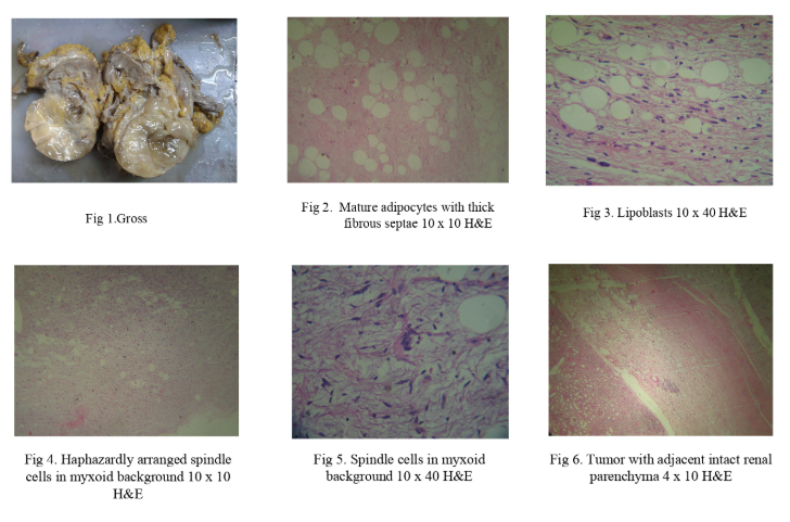
Gross: [Fig 1] showed a grey brown mass measuring 23x15x12cm with bosselated outer surface . Cut section revealed a tumour measuring 10x9cm with yellowish brown, soft glistening cut surface with compressed adjacent renal parenchyma and pelvis.
Microscopy- Showed areas of adipose tissue with atypical adipocytes few of them resembled lipoblasts[Fig 3]. Also seen were scattered hyperchromatic as well as multinucleated stromal cells within thick fibrous septa[Fig 2&5].
Areas with spindle cells arranged haphazardly with intervening myxoid material resembling myxofibrosarcoma were seen[Fig 4].
Renal parenchyma appeared completely delineated from the tumor and showed intact glomeruli and tubules[Fig 6] With the above features a diagnosis of De-differentiated Liposarcoma was made.
Discussion:
Liposarcoma arising in extremities or retroperitoneum affects middle-aged and old patients.[1,4]
Liposarcoma of kidney is a rare tumor with only few well-documented reports in the literature. [3,5]
Dedifferentiated liposarcoma, with its high-grade histology exhibits an aggressive clinical course.[1]
An important differential diagnosis is AML because both are large fat-containing lesions.[1,2]
Liposarcomas arise in perirenal fat within Gerota’s fascia or capsule and they displace, compress, and distort the kidney but usually do not invade the adjacent renal parenchyma.[5]
Conclusion
It is difficult to establish the exact origin of the tumor radiologically as Liposarcomas do not cause any defect in the renal parenchyma and interface between tumor and kidney is smooth. Hence histology becomes the gold standard for diagnosis [1].
References:
Charu shastri, Jatinder Kumar, Sushila Jaiswal and Anil Mandhani, Renal dedifferentiated liposarcoma with intracaval tumor thrombus: A rare case, Indian J Urol. 2012 Apr-Jun; 28(2): 208–210.
Fletcher CD, Unni KK, Mertens F. Pathology and Genetics of Tumours of soft tissue and bone. Lyon: IARC Press; 2002. World Health Organization Classification of Tumors; pp. 40–4. (227-32).
Matsushita M, Ito A, Ishidoya S, Endoh M, Moriya T, Arai Y. Intravenous extended liposarcoma arising from renal sinus. Int J Urol. 2007;14:769–70.
Histologic Subtype and Margin of Resection Predict Pattern of Recurrence and Survival for Retroperitoneal Liposarcoma. Ann Surg. 2003;238:358–71.
Diago A.L.Bader, Luis A.B.Peres, Sergio L. Bader, Renal Liposarcoma, International Braz J Urol Vol. 30(3): 214-215, May-June 2004
Leave a Reply